Obscure Gastrointestinal blood loss
and iron deficiency anaemia
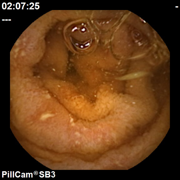
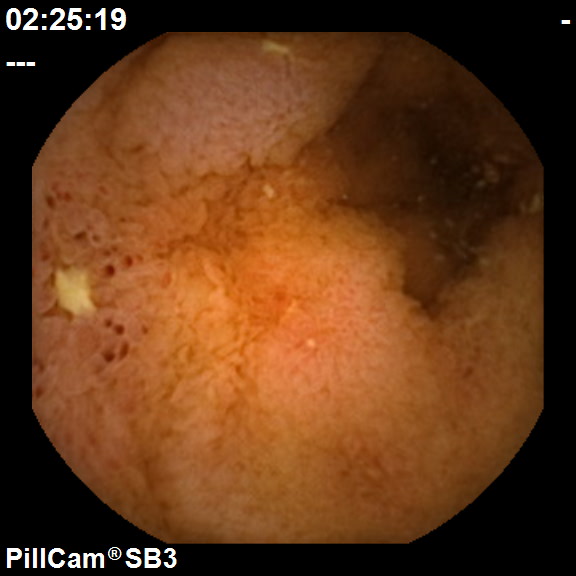
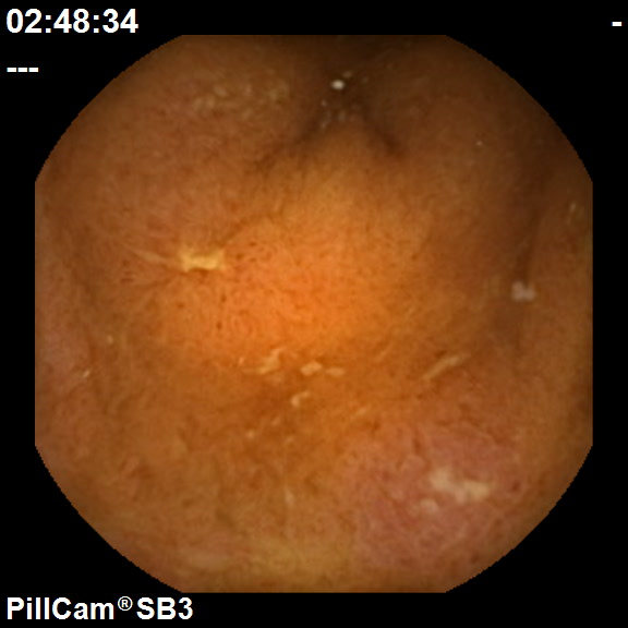
Obscure GastrointestinaI blood loss often manifests as melaena (black, tarry stools due to altered blood) or iron deficiency anaemia. When gastroscopy and colonoscopy has failed to find a source of bleeding, capsule endoscopy is the best way to visualise the hidden parts of the small intestine and exclude a serious cause.


Click on each image to view:
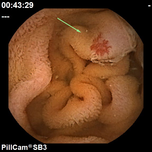
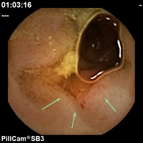
Angioectasia
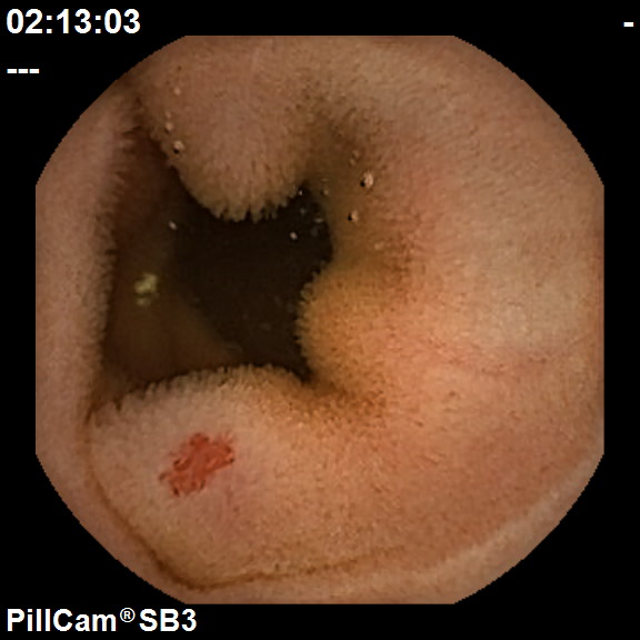
Angioectasia 1